2021: Clubroot on Canola: Survey & Quantification of Resting Spores of Plasmodiophora brassicae from Field Collected Soil Samples in North Dakota
(Research Report, Langdon REC, December 2021)Take Home Message: An ongoing clubroot survey for over six years in various counties of North Dakota indicates a threat to the canola crop if proper attention is not given towards longer crop rotations (1 in 3 years). In addition, growers should consider growing an available clubroot resistant canola variety in endemic areas and following proper equipment sanitation. Cleaning equipment thoroughly after working in a clubroot infected field is highly recommended since the primary mechanism of spread between fields is the movement of infested soil on farm equipment.
Survey Procedure: The survey involved three components: 1. visual survey, 2. soil sampling, and 3. molecular quantification of resting spores of the clubroot pathogen.
Components 1&2. Visual survey and soil sampling: Clubroot scouting was done visually by inspecting canola crop roots. The disease survey was conducted in over 40 counties in North Dakota. One field in every 5000 acres was targeted for scouting in each county. Soil samples were collected from fields to determine the pH of the soil and the number of resting spores per gram of soil. A minimum of 3-10 fields per county were targeted for scouting.
The survey was done in two phases.
1st phase: at flowering (10% of flowering onwards)
Plants were sampled from distinct stunted patches or prematurely senescing plants in the field during the growing season. Patches visible from the edge of the field were checked by digging and observing the roots for symptoms of clubroot and soil samples were collected from those spots.
2nd phase: after swathing
Scouting at swathing was based on the methodology followed in Canada by the Alberta Agricultural and Rural Development (AARD) for their annual clubroot disease survey. Reports of AARD indicated that the probability of finding clubroot was higher if scouted at the field entrances. Hence, the survey was done starting from the main entrances/approaches in each field the survey group visited and walked in a “W” pattern stopping at 5 spots and uprooting 10 consecutive stems from the ground at each spot. Each sampling point was separated by 100 meters or 328 feet. Roots of 50 stems were evaluated for the presence of clubroot and incidence. After removing excess soil, roots were visually examined for the presence of galls. At sample sites where infection was observed or suspected, root specimens with galls, along with soil, were double bagged and labeled with the field location. Infected roots and soil samples from all the fields surveyed were collected and a representative sample was submitted to Dr. Zhaohui Liu’s laboratory for molecular quantification of resting spores per gram of soil. An additional half-pound of soil was sent to the NDSU Soil Testing Laboratory for pH determination.
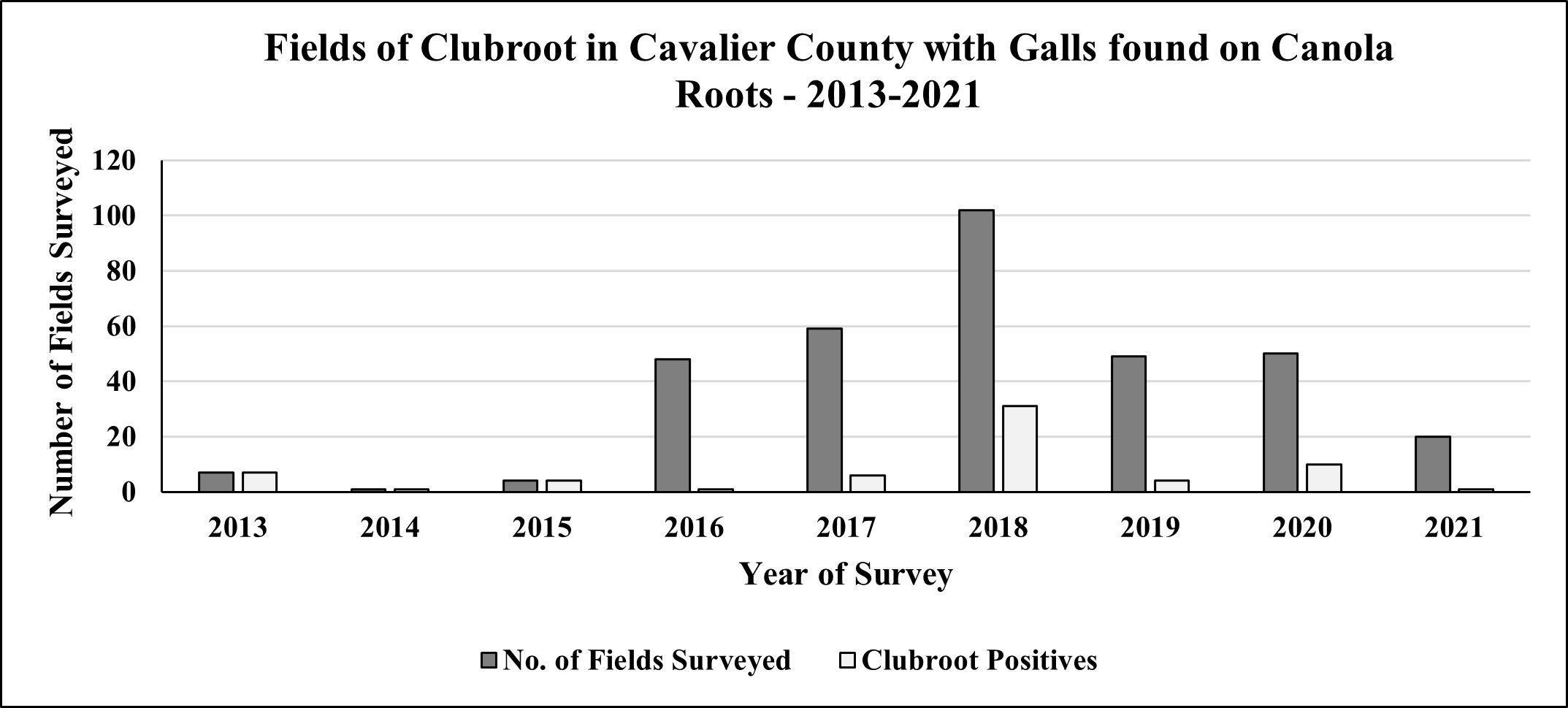
Results: Over 49 counties in North Dakota were surveyed in 2021 for visual symptoms of galls on canola roots. Clubroot galls on canola roots were only found in 1 out of 20 fields surveyed in Cavalier County (Figure 1).
Figure 1: Fields surveyed from 2013 to 2021 for prevalence of clubroot in Cavalier County, North Dakota.
Component 3. Molecular detection of soil samples to quantify Plasmodiophora brassicae (the clubroot pathogen) resting spores:
Since a large number of samples were submitted for quantification in 2020, results were delayed and not available until March 2021. They are presented in this report. Likewise, approximately 400 soil samples have been submitted for the 2021 season from 49 counties and are awaiting results which will be presented in the 2022 annual report.
The main objective of this procedure is to quantify resting spores of the clubroot pathogen from the soil and inform growers prior to the occurrence of visible gall symptoms in canola.
Results from molecular assays on soil samples in 2020: The molecular assays indicated the clubroot pathogen resting spores have been found in 26 out of 44 counties (Figure 2) surveyed. Although, there were no visible symptoms observed when the roots were uprooted. Quantified resting spores of P. brassicae from those samples ranged from 500 to 40,000,000 spores per gram of soil (minimum detection limit of the assay being 10 resting spores/g of soil). Lack of visible galls in the surveyed fields but positive in the molecular soil quantification assay indicate either the resting spore population may not have reached required spores per gram of soil in acidic soils to show galls or the pH of soil is basic (Figure 3). In general, clubroot infections are expressed on canola plants where soil resting spore population total about 80,000 spores per gram of soil (Canadian Research). These results indicate that there is a need for continuous annual monitoring.
Notice: Growers who are curious about the presence of clubroot/resting spores in their field(s) are encouraged to contact Dr. Venkat Chapara at the Langdon REC (701-256-2582), NDSU Cavalier County Extension Office (701-256-2560), or NDSU Extension (701-231-8363).
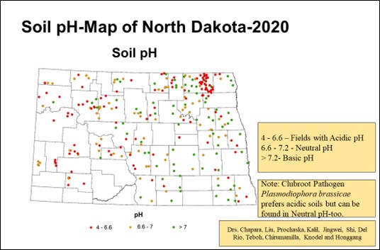
Figure 2: Map of counties in North Dakota indicating Plasmodiophora brassicae (the clubroot pathogen) resting spores detected by molecular assays in the soil samples submitted in 2020.
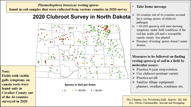
Note: The green dot indicates zero, yellow dot indicates range of 1-80,000 and the red dot indicates more than 80,000 resting spores per gram of soil.
Figure 3: Map of counties in North Dakota indicating pH ranges detected by soil assays in the soil samples submitted in 2020.