Title

Soybean Disease Diagnostic Series

(PP1867, Reviewed Jan. 2024)
Summary

This series aids in disease identification.

Lead Author
Lead Author:
Samuel Markell, Associate Professor and Extension Specialist, Department of Plant Pathology, North Dakota State University
Other Authors

Dean Malvick, Professor and Extension Specialist, Department of Plant Pathology, University of Minnesota

Availability
Availability:
Web only
Publication Sections
Row of Soybeans
Photo Credit:
Sam Markell, NDSU

Root Diseases

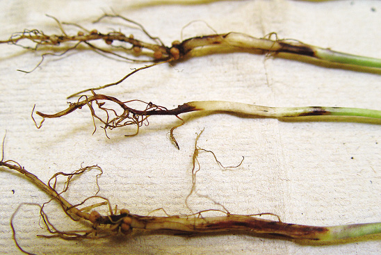
Fusarium root rot

Fusarium solani, F. oxysporum, F. tricinctum and other Fusarium species (fungi)

FIGURE 1 – Dark brown/black discoloration of roots
Photo Credit:
B. Nelson, NDSU
FIGURE 1 – Dark brown/black discoloration of roots
FIGURE 2 – Root rot and dieback of tap root
Photo Credit:
Giesler, Univ. of Nebraska
FIGURE 2 – Root rot and dieback of tap root
FIGURE 3 – Seedling damping-off
Photo Credit:
J. Bienapfl, Univ. of Minnesota
FIGURE 3 – Seedling damping-off
FIGURE 4 – Plant chlorosis
Photo Credit:
D. Mueller, Iowa State Univ.
FIGURE 4 – Plant chlorosis

AUTHORS: Dean Malvick and Sam Markell

SYMPTOMS

• Brown to black discoloration and rot of roots, especially the taproot
• Plant stunting and yellowing of leaves may occur if root rot is severe
• Seedling damping-off

FACTORS FAVORING DEVELOPMENT

• Soil compaction
• Presence of soybean cyst nematode (SCN)
• Plant stress
• Drought

IMPORTANT FACTS

• Disease can infect and kill seedlings and damage older plants
• Fusarium survives for long periods in soil
• Dry edible beans, corn and pulse crops are hosts
• Fusarium is dispersed with soil (on equipment, in water, by wind, etc.)
• Management: improve soil drainage, reduce compaction, fungicide seed treatments
• Commonly confused with other roots rots, SCN and iron deficiency chlorosis


Phytophthora root and stem rot

Phytophthora sojae (oomycete)

FIGURE 1 – Close-up of lower stem lesion
Photo Credit:
D. Malvick, Univ. of Minnesota
FIGURE 1 – Close-up of lower stem lesion
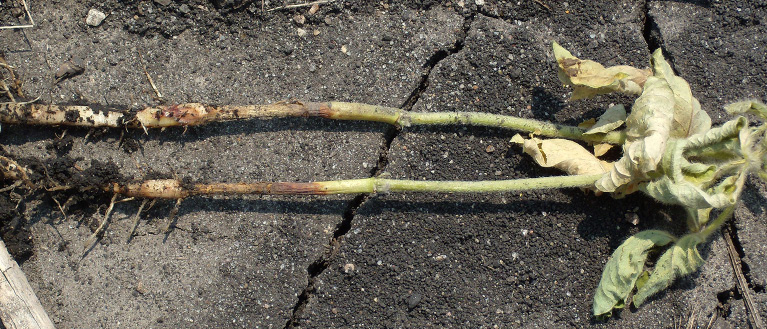
FIGURE 2 – Lower stem lesion and wilting
Photo Credit:
D. Malvick, Univ. of Minnesota
FIGURE 2 – Lower stem lesion and wilting
FIGURE 3 – Severe infection in field
Photo Credit:
A. Dorrance, Ohio State Univ.
FIGURE 3 – Severe infection in field

AUTHORS: Sam Markell, Dean Malvick and Berlin Nelson

SYMPTOMS

• Seeds may rot and/or seedlings may die before or after emergence (damping-off)
• Mid- to late-season symptoms include a chocolate brown stem lesion extending up from the soil line
• Leaf chlorosis, necrosis and plant wilting can develop
• Frequently occurs in patches of fields and low areas

FACTORS FAVORING DEVELOPMENT

• Excessive moisture, saturated soil and/or flooding, especially early in the season
• Poorly drained, heavy clay or compacted soils
• Short/no crop rotation

IMPORTANT FACTS

• Can cause significant yield loss
• The pathogen is specific to soybeans
• Management tools available include genetic resistance and seed treatments
• The pathogen has many pathotypes and many can overcome Rps genetic resistance
• Commonly confused with other root rot diseases

Pythium root rot

Pythium ultimum and other Pythium species (oomycete)

FIGURE 1 – Seed rotting due to Pythium
Photo Credit:
D. Malvick, Univ. of Minnesota
FIGURE 1 – Seed rotting due to Pythium
FIGURE 2 – Damping-off of seedlings
Photo Credit:
M. Chilvers, Michigan State Univ.
FIGURE 2 – Damping-off of seedlings
FIGURE 3 – Light brown infected roots
Photo Credit:
A. Robertson, Iowa State Univ.
FIGURE 3 – Light brown infected roots

AUTHORS: Dean Malvick and Sam Markell

SYMPTOMS

• Soft, slimy rot of seeds in ground
• Light brown rot of outer part of roots (cortex)
• Damping-off of seedlings

FACTORS FAVORING DEVELOPMENT

• Persistent wet soil after planting
• Soil compaction, heavy soil with high clay content
• Slow growth of seedlings and plant stress

IMPORTANT FACTS

• Pythium survives for years in soil
• Pathogen causes most damage to seeds and seedlings, but may damage roots of older plants
• Dry edible beans, corn and other crops can be hosts
• Pythium is dispersed with soil (on equipment, in water, by wind, etc.)
• Management: improve soil drainage, reduce compaction, some seed treatments
• Symptoms on seed and seedlings very similar to Phytophthora damage
• Commonly confused with other seedling diseases

Rhizoctonia root rot

Rhizoctonia solani (fungus)

FIGURE 1 – Rusty-brown lesions on soybean stems
Photo Credit:
D. Malvick, Univ. of Minnesota
FIGURE 1 – Rusty-brown lesions on soybean stems
FIGURE 2 – Soybean seedlings with girdled stems
Photo Credit:
D. Malvick, Univ. of Minnesota
FIGURE 2 – Soybean seedlings with girdled stems
FIGURE 3 – Seedlings dying in a row
Photo Credit:
D. Malvick, Univ. of Minnesota
FIGURE 3 – Seedlings dying in a row

AUTHORS: Dean Malvick and Sam Markell

SYMPTOMS

• Rusty-brown, dry, sunken lesions on lower parts of stems
• Dark brown lesions than girdle the stems near the soil
• Plants stunted, yellow, and wilting

FACTORS FAVORING DEVELOPMENT

• Warm and moist soil while plants are in early vegetative stages
• Delayed planting in spring due to rain
• High soil organic matter
• Plant stress due to physical or chemical/herbicide injury

IMPORTANT FACTS

• Dry edible beans, corn, sugar beet and pulse crops can be hosts
• Causes most damage to plants in early vegetative growth stages
• Rhizoctonia is dispersed with soil (on equipment, in water, by wind, etc.)
• Management: some seed treatments, crop rotation, tillage, early planting
• Soybean varieties vary in susceptibility to Rhizoctonia
• Commonly confused with other roots rots 

Seed and seedling disease complex

Pythium (oomycete), Phytophthora (oomycete), Rhizoctonia (fungus), Fusarium (fungus)

FIGURE 1 – Seedling decomposing due to infection
Photo Credit:
D. Malvick, Univ. of Minnesota
FIGURE 1 – Seedling decomposing due to infection
FIGURE 2 – Pre-emergence death of seedling
Photo Credit:
D. Malvick, Univ. of Minnesota
FIGURE 2 – Pre-emergence death of seedling
FIGURE 3 – Post-emergence death of seedling
Photo Credit:
D. Malvick, Univ. of Minnesota
FIGURE 3 – Post-emergence death of seedling

AUTHORS: Dean Malvick and Sam Markell

SYMPTOMS

• Rotting of seed in the ground
• Pre-emergence death
• Post-emergence death and stunting
• Soft, tan roots with intact steel (Pythium and Phytophthora)
• Dark brown discoloration and rot of tap roots (Fusarium)
• Rusty-brown lesions on stem and roots (Rhizoctonia)

FACTORS FAVORING DEVELOPMENT

• Wet soil
• Delayed germination and growth
• Poor-quality seed
• Plant stress

IMPORTANT FACTS

• Pathogens are favored by different conditions for infection and disease development
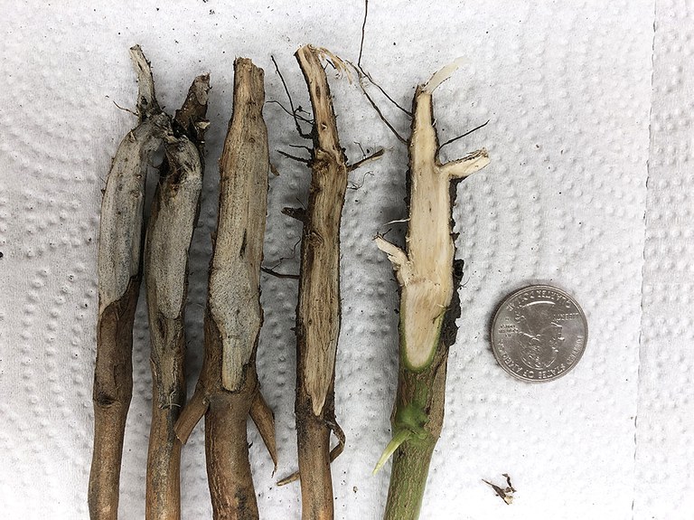
• Multiple pathogens often infect seeds/seedlings
• Difficult to diagnose principle pathogen
• Symptoms can look similar for different pathogens, but management may differ
• Management options: soil drainage, tillage, varieties with resistance, seed treatments
• Seed treatment ingredients vary in efficacy for different pathogens
• Commonly confused with water damage

Soybean cyst nematode (SCN)

Heterodera glycines (plant parasitic nematode)

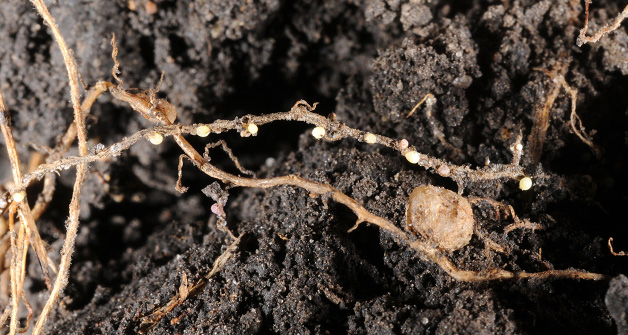
FIGURE 1 – White SCN females (cysts) and a nodule on soybean roots
Photo Credit:
S. Markell, NDSU
FIGURE 1 – White SCN females (cysts) and a nodule on soybean roots
FIGURE 2 – Mature brown cysts
Photo Credit:
S. Markell, NDSU
FIGURE 2 – Mature brown cysts
FIGURE 3 – Yellowed areas near field entrance caused by severe SCN
Photo Credit:
S. Markell, NDSU
FIGURE 3 – Yellowed areas near field entrance caused by severe SCN

AUTHORS: Sam Markell and Dean Malvick

SYMPTOMS

• Small (approximately 1/32 inch) lemon-shaped female worms (cysts) on roots
• Cysts’ color ranges from cream to dark brown
• Above-ground symptoms often are absent
• Soybeans can become stunted and yellow

FACTORS FAVORING DEVELOPMENT

• Short crop rotation with soybeans and/or dry edible beans
• High soil pH, light soil texture
• Dry growing seasons

IMPORTANT FACTS

• SCN is the most yield-limiting disease in the U.S.
• SCN can make other diseases (sudden death syndrome, brown stem rot) worse
• Soybeans, dry edible beans and several weeds are hosts
• SCN is dispersed with soil (on equipment, in water, by wind, etc.)
• Soil testing is the most reliable way to determine if you have SCN
• Management tools available include crop rotation, variety resistance and possibly seed treatments
• SCN is overcoming PI88788 genetic resistance
• Commonly confused with roots rots, iron deficiency chlorosis, other abiotic stress

Sudden death syndrome (SDS)

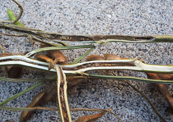
Fusarium virguliforme (fungus)

FIGURE 1 –  Internal stem browning
Photo Credit:
D. Malvick, Univ. of Minnesota
FIGURE 1 – Internal stem browning
FIGURE 1 – Early chlorotic blotches between veins
Photo Credit:
D. Malvick, Univ. of Minnesota
FIGURE 1 – Early chlorotic blotches between veins
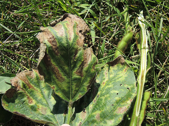
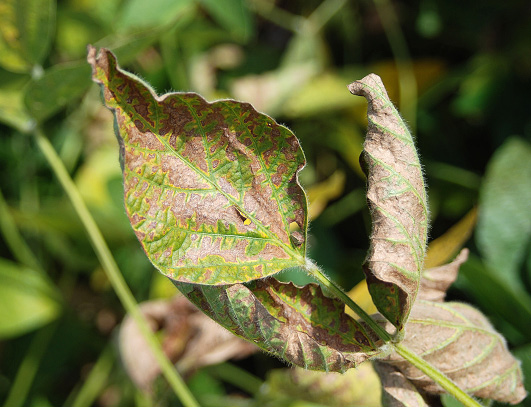
FIGURE 2 – Severe leaf symptoms
Photo Credit:
D. Malvick, Univ. of Minnesota
FIGURE 2 – Severe leaf symptoms
FIGURE 3 – Severe infection in a patch in a field
Photo Credit:
D. Malvick, Univ. of Minnesota
FIGURE 3 – Severe infection in a patch in a field
FIGURE 4 – Root rot and blue fungal growth on root
Photo Credit:
D. Malvick, Univ. of Minnesota
FIGURE 4 – Root rot and blue fungal growth on root

AUTHORS: Dean Malvick and Sam Markell

SYMPTOMS

• Yellow and brown discoloration between leaf veins
• Tan stem under the epidermis near the soil line, while pith remains white
• Symptoms develop in plant seed fill stages (August)
• Leaflets can fall and petioles remain attached to plant
• Root rot and occasionally blue fungal growth on root

FACTORS FAVORING DEVELOPMENT

• Wet soil for two to four weeks after planting
• Field history of SDS
• Compacted soil, poor drainage
• Periodic heavy rain and moist soil through mid-Aug.
• High soybean cyst nematode populations

IMPORTANT FACTS

• Common in southern and central Minnesota, less common (but increasing) in northern Minnesota and North Dakota
• Pathogen can cause root rot of dry edible bean and some other legumes
• Pathogen dispersed with soil (on equipment, in water, by wind, etc.)
• Management options include varieties with resistance to SDS and seed treatments
• Commonly confused with brown stem rot

Stem Diseases

Anthracnose

Colletotrichum truncatum or other Colletotrichum species (fungi)

FIGURE 1 – Irregular-shaped blotches on stem
Photo Credit:
D. Malvick, Univ. of Minnesota
FIGURE 1 – Irregular-shaped blotches on stem
FIGURE 2 – Abundant black fungal growths of irregular arrangement and size
Photo Credit:
S. Markell, NDSU
FIGURE 2 – Abundant black fungal growths of irregular arrangement and size
FIGURE 3 – Close-up appearance of fungal growths
Photo Credit:
S. Markell, NDSU
FIGURE 3 – Close-up appearance of fungal growths

AUTHORS: Sam Markell and Dean Malvick

SYMPTOMS

• Symptoms usually not seen until plants reach maturity
• Dark brown patches may appear on stems, pods and petioles
• Small black fungal spots develop in irregular patterns on stems, pods and petiole
• Black, infected areas covered with tiny black spines (setae) that can be seen with a 10X hand lens

FACTORS FAVORING DEVELOPMENT

• Warm and wet/humid weather
• Planting infected seed
• Short/no crop rotation

IMPORTANT FACTS

• A common late-season disease that rarely causes significant yield loss
• Pathogen(s) have a wide host range and may infect other legume crops and weeds
• Pathogen can be seedborne
• Commonly confused with pod and stem blight, stem canker, charcoal rot


Brown stem rot (BSR)

Cadophora gregata (fungus)

FIGURE 1 – Light brown discoloration in pith and leaf
Photo Credit:
D. Malvick, Univ. of Minnesota
FIGURE 1 – Light brown discoloration in pith and leaf
FIGURE 2 – Dark brown discoloration in pith of stem
Photo Credit:
D. Malvick, Univ. of Minnesota
FIGURE 2 – Dark brown discoloration in pith of stem
FIGURE 3 – Brown and yellow discoloration between veins
Photo Credit:
D. Malvick, Univ. of Minnesota
FIGURE 3 – Brown and yellow discoloration between veins
FIGURE 4 – Symptoms on leaves of whole plant
Photo Credit:
D. Malvick, Univ. of Minnesota
FIGURE 4 – Symptoms on leaves of whole plant

AUTHORS: Dean Malvick and Sam Markell

SYMPTOMS

• Brown pith in stem, especially in lower stem
• Brown and yellow discoloration between leaf veins may be present
• Symptoms commonly develop in mid-August

FACTORS FAVORING DEVELOPMENT

• Short/no crop rotation
• Wet and cool weather in July/August
• History of disease in a field
• Susceptible soybean varieties
• Presence of soybean cyst nematode

IMPORTANT FACTS

• Stems should be longitudinally split to identify BSR
• Commonly confused with sudden death syndrome
• Pathogen overwinters/survives in infected soybean stems
• BSR pathogen dispersed with soil (on equipment, in water, by wind, etc.)
• Soybean is only known definite host
• Two pathogen types: type A causes leaf and stem symptoms; type B primarily internal stem symptoms only
• Type B more common in our region
• Management options: crop rotation and varieties resistant to BSR

Charcoal rot

Macrophomina phaseolina (fungus)

FIGURE 1 – Large patch of soybean with charcoal rot
Photo Credit:
B. Nelson, NDSU
FIGURE 1 – Large patch of soybean with charcoal rot
FIGURE 2 – Patch of wilting soybeans
Photo Credit:
B. Nelson, NDSU
FIGURE 2 – Patch of wilting soybeans
FIGURE 3 – Infected (L and C) and healthy soybean (R)
Photo Credit:
B. Nelson, NDSU
FIGURE 3 – Infected (L and C) and healthy soybean (R)
FIGURE 4 – External gray lesion peeling away, revealing profuse "charcoal" sclerotia
Photo Credit:
S. Markell, NDSU
FIGURE 4 – External gray lesion peeling away, revealing profuse "charcoal" sclerotia

AUTHORS: Sam Markell and Dean Malvick

SYMPTOMS

• Symptoms usually not apparent until flowering or later
• Taproot and lower stem may appear gray/silver
• Numerous black fungal specks (microsclerotia) under epidermis give a “charcoal” appearance
• Premature death with wilted leaves attached
• Frequently occurs in patches in fields

FACTORS FAVORING DEVELOPMENT

• Hot temperatures
• Drought stress

IMPORTANT FACTS

• Yield loss may occur in hot, dry growing seasons
• Disease typically most severe in drought-prone areas of fields
• Very wide host range, which includes corn, sunflower, other legume crops and weeds
• Commonly confused with anthracnose, Phytophthora stem rot, pod and stem blight, stem canker

Pod and stem blight/Phomopsis seed decay

Diaporthe longicolla (fungi) and D. sojae

FIGURE 1 – Linear rows of raised black dots
Photo Credit:
D. Malvick, Univ. of Minnesota
FIGURE 1 – Linear rows of raised black dots
FIGURE 2 – Infected (L) and healthy (R) plants
Photo Credit:
D. Malvick, Univ. of Minnesota
FIGURE 2 – Infected (L) and healthy (R) plants

AUTHORS: Sam Markell and Dean Malvick

SYMPTOMS

• Small, raised black dots (pycnidia) arranged in distinct rows on stem, pods and petioles
• Tops of plants may discolor and die, leading to plant death
• Symptoms often not apparent until plants near maturity
• Wavy, black zone lines inside infected stems and roots may occur (see stem canker)
• Seed may be cracked, shriveled, moldy and have poor germination

FACTORS FAVORING DEVELOPMENT

• Warm and humid weather
• Short/no crop rotation
• Planting infected seed
• Delayed harvest due to wet weather

IMPORTANT FACTS

• Yield loss and reduction in seed quality may occur
• Host range includes dry edible bean and dry edible pea
• Pathogen survives in crop residue and seed
• Pathogen is widespread, even in apparently healthy plants
• Black dots arranged in rows are diagnostic (anthracnose and charcoal rot dots are random)
• Commonly confused with anthracnose, charcoal rot and stem canker

Stem canker

Diaporthe caulivora (northern stem canker) and D. aspalathi (southern stem canker) (fungi)

FIGURE 1 – Reddish-brown stem canker lesion
Photo Credit:
D. Malvick, Univ. of Minnesota
FIGURE 1 – Reddish-brown stem canker lesion
FIGURE 2 – Sunken canker on lower stem of mature plant
Photo Credit:
S. Markell, NDSU
FIGURE 2 – Sunken canker on lower stem of mature plant
FIGURE 3 – Zone lines beneath epidermis of sunken canker
Photo Credit:
S. Markell, NDSU
FIGURE 3 – Zone lines beneath epidermis of sunken canker


AUTHORS: Sam Markell and Dean Malvick

SYMPTOMS

• Reddish-brown lesions on the lower stem starting at branch points/nodes
• Lesions expand and may become sunken cankers
• Tiny black fungal structures may be produced on lesions
• Narrow black “zone lines” sometimes under epidermis, but importantly, zone lines also are associated with pod and stem blight

FACTORS FAVORING DEVELOPMENT

• Extended periods of wet weather (one to four days) with moderate temperatures (70 to 85 F)
• Short/no crop rotation in fields with history of disease
• Reduced tillage

IMPORTANT FACTS

• Yield loss can occur if disease is widespread in a field and kills plants before pod fill
• Management tools include crop rotation (wheat, corn, etc.), resistant varieties and foliar fungicides
• Commonly confused with anthracnose, charcoal rot, late-season Phytophthora root rot, and pod and stem blight

White mold (Sclerotinia stem rot)

Sclerotinia sclerotiorum (fungus)

FIGURE 1 – Lesions with white mold and sclerotia
Photo Credit:
D. Malvick, Univ. of Minnesota
FIGURE 1 – Lesions with white mold and sclerotia
FIGURE 2 – Severe white mold infection
Photo Credit:
M. Gilley, NDSU
FIGURE 2 – Severe white mold infection
FIGURE 3 – Black sclerotia among shriveled seeds
Photo Credit:
S. Markell, NDSU
FIGURE 3 – Black sclerotia among shriveled seeds
FIGURE 4 – Apothecia
Photo Credit:
S. Markell, NDSU
FIGURE 4 – Apothecia

AUTHORS: Sam Markell and Dean Malvick

SYMPTOMS

• Stem lesions begin as water-soaked spots near nodes
• Lesions enlarge, fluffy white fungal growth develops on moist stems
• Infected stems become bleached white and may shred
• Hard black structures (sclerotia) form on and in infected tissue

FACTORS FAVORING DEVELOPMENT

• Wet soils prior to and during soybean flowering
• Frequent wetness (rain, fog, heavy dew) and cool temperatures during bloom
• Dense plant canopy, high fertility, high plant populations
• History of white mold in field

IMPORTANT FACTS

• Severe yield losses can occur when July and early August are cool and wet
• Many broadleaf crops and weeds are hosts
• Pathogen survives in soil for many years as sclerotia
• Sclerotia produce apothecia (about 1/4-inch mushrooms), which produce ascospores that initiate infection
• Management options: partially resistant varieties, increase plant spacing and fungicides
• Apothecia commonly confused with bird’s nest fungi

Leaf Diseases

Bacterial blight

Pseudomonas savastanoi pv. glycinea (bacteria)

FIGURE 1 – Brown angular lesions with bright yellow halos
Photo Credit:
S. Markell, NDSU
FIGURE 1 – Brown angular lesions with bright yellow halos
FIGURE 2 – Coalescing lesions and leaf tattering
Photo Credit:
S. Markell, NDSU
FIGURE 2 – Coalescing lesions and leaf tattering
FIGURE 3 – Magnified lesions
Photo Credit:
S. Markell, NDSU
FIGURE 3 – Magnified lesions

AUTHORS: Sam Markell and Dean Malvick

SYMPTOMS

• Typically observed first in upper canopy in July
• Small, water-soaked and angular leaf lesions
• Lesion centers turn brown and are surrounded by a bright yellow halo
• Lesions often coalesce and leaves will tatter
• Often widespread distribution in field

FACTORS FAVORING DEVELOPMENT

• Cool temperatures, frequent rains and thunderstorms
• Weather that damages plant tissue (hail, high winds, etc.)
• Short/no crop rotation
• Planting infected seed

IMPORTANT FACTS

• Widespread but rarely economically important
• Lesions may occur on stem, petiole and pod
• Pathogen survives and can be spread with seed and infested crop residue
• Fungicides are not effective
• Commonly confused with Septoria brown spot, bacterial pustule, downy mildew

Bacterial pustule

Xanthomonas axonopodis pv. glycines (bacteria)

FIGURE 1 – Lesions with chlorotic halos on upper side of leaf
Photo Credit:
FIGURE 1 – Lesions with chlorotic halos on upper side of leaf
FIGURE 2 – Lesions and pustules on underside of leaf
Photo Credit:
S. Markell, NDSU
FIGURE 2 – Lesions and pustules on underside of leaf
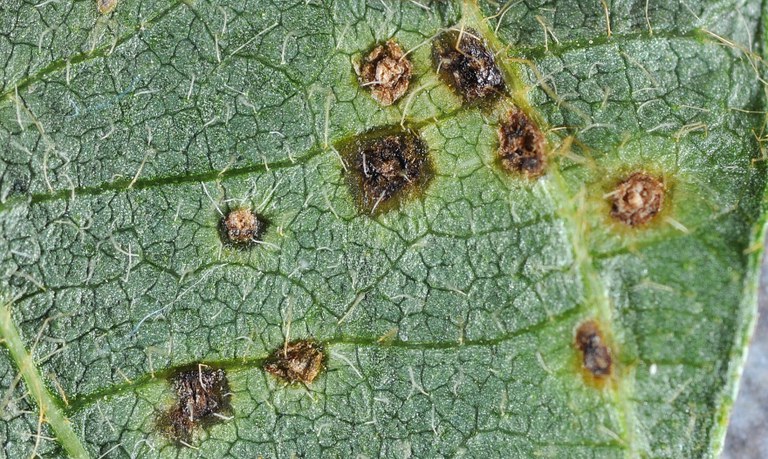
FIGURE 3 – Pustules (approximately 5 to 10X)
Photo Credit:
S. Markell, NDSU
FIGURE 3 – Pustules (approximately 5 to 10X)

AUTHORS: Sam Markell and Dean Malvick

SYMPTOMS

• Lesions begin as small (1/16 to 1/4 inch) light green specks with yellow halos
• Lesion centers turn brown
• Raised pustules appear in lesions

FACTORS FAVORING DEVELOPMENT

• Wet and rainy weather
• Prolonged humid conditions
• Warm to hot temperatures

IMPORTANT FACTS

• Sporadic in Minnesota and North Dakota
• Unlikely to cause yield loss
• Can be mistaken for soybean rust, a disease that has not occurred in Minnesota or North Dakota
• Commonly confused with bacterial blight or Septoria brown spot

Bean pod mottle virus

FIGURE 1 – Light green to yellow leaf mottling
Photo Credit:
A. Dorrance, Ohio State Univ.
FIGURE 1 – Light green to yellow leaf mottling
FIGURE 2 – Wrinkling, puckering and light green mottling
Photo Credit:
A. Dorrance, Ohio State Univ.
FIGURE 2 – Wrinkling, puckering and light green mottling
FIGURE 3 – Leaf puckering and wrinkling
Photo Credit:
Univ. of Illinois
FIGURE 3 – Leaf puckering and wrinkling


AUTHORS: Dean Malvick and Sam Markell

SYMPTOMS

• Green to yellow mottling of young leaves
• Leaves may become puckered and wrinkled
• Symptoms may not be visible during high temperatures or after pod set
• Seed may become mottled with dark stains

FACTORS FAVORING DEVELOPMENT

• High populations of bean leaf beetle (or other beetles) early in the season
• Cool weather

IMPORTANT FACTS

• Not thought to be common in Minnesota and North Dakota
• Virus can be transmitted by bean leaf beetle or other leaf feeding beetles
• Infection primarily occurs early in the season
• Host range includes dry edible bean, clovers and other legumes
• Management options: delay planting, seed applied insecticides
• Commonly confused with other viruses and possibly herbicide injury

Cercospora leaf blight

Cercospora kikuchii (fungus)

FIGURE 1 – Purple discoloration of leaf
Photo Credit:
A.K. Chanda, Univ. of Minnesota
FIGURE 1 – Purple discoloration of leaf
FIGURE 2 – Bronze discoloration and death of leaf tissue
Photo Credit:
A.K. Chanda, Univ. of Minnesota
FIGURE 2 – Bronze discoloration and death of leaf tissue
FIGURE 3 – Purple seed stain
Photo Credit:
Univ. of Illinois
FIGURE 3 – Purple seed stain

AUTHORS: Dean Malvick and Sam Markell

SYMPTOMS

• Purple to bronze discoloration of upper leaf surfaces
• Red-brown spots on both leaf surfaces
• Large necrotic areas can develop on leaves, followed by leaf drop
• Seed coats can develop purple discoloration

FACTORS FAVORING DEVELOPMENT

• High humidity and warm temperatures
• Lack of crop rotation

IMPORTANT FACTS

• Disease is more common and severe in southern U.S.
• Pathogen overwinters on infested soybean debris and seed
• Plants susceptible from flowering to maturity
• Management options: pathogen-free seed, susceptible varieties, crop rotation and fungicides
• Commonly confused with sunscald

Downy mildew

Peronospora manshurica (Oomycete)

FIGURE 1 – Discrete lesions top side of leaf
Photo Credit:
S. Markell, NDSU
FIGURE 1 – Discrete lesions top side of leaf
Downy Mildew
Photo Credit:
S. Markell, NDSU
A.
FIGURE 2 – Fungal growth opposite lesions on underside of leaf (A) and the magnification of fungal growth (B)
Photo Credit:
S. Markell, NDSU
FIGURE 2 – Fungal growth opposite lesions on underside of leaf (A) and the magnification of fungal growth (B)
FIGURE 3 – Severe infection
Photo Credit:
D. Helland, Wilbur Ellis
FIGURE 3 – Severe infection

AUTHORS:Sam Markell and Dean Malvick

SYMPTOMS

• Lesions begin as discrete pale green to light yellow spots on top side of leaves
• Fluffy tan tufts of fungal growth occur opposite lesions on underside of leaves
• Lesions become brighter yellow and turn brown with age

FACTORS FAVORING DEVELOPMENT

• Frequent and prolonged periods of high humidity or free moisture (dew)
• Moderate temperatures
• Short crop rotation
• Planting infected seed or field history of downy mildew

IMPORTANT FACTS

• Yield loss thought to be rare; however, severe outbreaks have occurred in North Dakota and Minnesota
• Pathogen is specific to soybeans and will not cause downy mildew of other crops
• Can be confused with Septoria brown spot, powdery mildew and bacterial diseases

Frogeye leaf spot

Cercospora sojina (fungus)

FIGURE 1 – Lesions of various size and stages of development
Photo Credit:
S. Markell, NDSU
FIGURE 1 – Lesions of various size and stages of development
FIGURE 2 – Spots and patterns of lesion development on leaf
Photo Credit:
D. Malvick, Univ. of Minnesota
FIGURE 2 – Spots and patterns of lesion development on leaf
Frogeye leaf spot
Photo Credit:
S. Markell, NDSU
FIGURE 3 – Upper and underside of lesion with fuzzy gray mold.
Photo Credit:
S. Markell, NDSU
FIGURE 3 – Upper and underside of lesion with fuzzy gray mold.

 AUTHORS: Dean Malvick and Sam Markell

SYMPTOMS

• Brown leaf spots surrounded by a darker reddish-brown or purple ring
• Centers of spots become tan as they age and develop black specks
• Spots may coalesce, fall out and kill large parts of leaves

FACTORS FAVORING DEVELOPMENT

• Warm and humid weather
• Highly susceptible soybean varieties

IMPORTANT FACTS

• Disease more common in southern areas of the Midwest
• Pathogen overwinters in infected soybean residue and seed
• Management options: crop rotation, tillage and fungicides
• Pathogen resistance to strobilurin (QoI, FRAC 11) fungicides is common in Minnesota and North Dakota
• Commonly confused with bacterial blight, Phyllosticta leaf spot and Septoria brown spot

Phyllosticta leaf spot

Phyllosticta sojicola (fungi)

FIGURE 1 - Gray irregular-shaped lesions with dark, narrow margins
Photo Credit:
K. Bissonnette
FIGURE 1 - Gray irregular-shaped lesions with dark, narrow margins
FIGURE 2 - V-shaped lesion with small black specs (pycnidia)
Photo Credit:
K. Bissonnette
FIGURE 2 - V-shaped lesion with small black specs (pycnidia)
FIGURE 3 - Large lesion with numerous pycnidia
Photo Credit:
K. Bissonnette
FIGURE 3 - Large lesion with numerous pycnidia

AUTHORS: Sam Markell and Dean Malvick

SYMPTOMS

  • May occur anytime in growing season
  • Leaf lesions begin as pale green spots
  • Lesions become gray to tan with narrow dark purple to brown margin
  • Lesions may be oval, circular, irregular or V-shaped
  • Small black specs (pycnidia) may appear in lesion centers
  • Lesions also may occur on petioles, stems and pods

FACTORS FAVORING DEVELOPMENT

  • Cool temperatures
  • Wet conditions
  • Short crop rotations
  • Infected Seed

IMPORTANT FACTS

  • Minor soybean disease that rarely impacts yield
  • Appears to be uncommon in Minnesota and North Dakota
  • Pathogen survives in infested crop residue and seed
  • Commonly confused with frogeye leaf spot, bacterial blight, Septoria brown spot

Powdery mildew

Erysiphe diffusa and E. glycines (fungi)

FIGURE 1 – White tufts of fungal growth
Photo Credit:
D. Malvick, Univ. of Minnesota
FIGURE 1 – White tufts of fungal growth
FIGURE 2 – Severe infection covering leaf
Photo Credit:
D. Malvick, Univ. of Minnesota
FIGURE 2 – Severe infection covering leaf
FIGURE 3 – Infection spreading in hot spot
Photo Credit:
D. Malvick, Univ. of Minnesota
FIGURE 3 – Infection spreading in hot spot

AUTHORS: Sam Markell and Dean Malvick

SYMPTOMS

• Powdery white tufts of fungal growth on upper side of leaf
• Fungal growth may look like white flour sprinkled on the leaves
• Fungal growth can expand and may cover entire leaf surface
• Small black specs in growth may be observed late in season
• White fungal growth can be rubbed off leaf easily

FACTORS FAVORING DEVELOPMENT

• Temperatures from 64 to 75 F
• Low humidity with periods of limited leaf wetness
• Late-planted soybeans

IMPORTANT FACTS

• Yield loss is rare in Minnesota and North Dakota
• Usually occurs late in growing season
• The pathogen also may infect dry edible beans and field peas
• Commonly confused with downy mildew

Septoria brown spot

Septoria glycines (fungus)

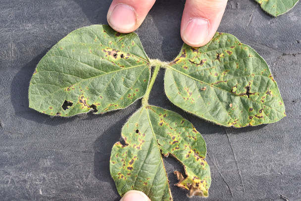
FIGURE 1 – Brown spots and chlorosis
Photo Credit:
D. Malvick, Univ. of Minnesota
FIGURE 1 – Brown spots and chlorosis
FIGURE 2 – Trifoliate with brown spots and chlorosis
Photo Credit:
D. Malvick, Univ. of Minnesota
FIGURE 2 – Trifoliate with brown spots and chlorosis
FIGURE 3 – Common pattern of symptoms developing in lower canopy
Photo Credit:
D. Malvick, Univ. of Minnesota
FIGURE 3 – Common pattern of symptoms developing in lower canopy

AUTHORS: Dean Malvick and Sam Markell

SYMPTOMS

• Dark brown spots (less than 1/8 inch diameter)
• Brown spots coalesce into large brown areas
• Irregular brown and yellow patches on one side of leaf
• Symptoms first develop in lower part of plant, then move up

FACTORS FAVORING DEVELOPMENT

• Wet and warm weather
• High plant density
• Continuous soybean planting
• Minimum tillage

IMPORTANT FACTS

• Typically does not cause yield loss
• Under severe conditions, defoliation and yield loss can occur
• Pathogen survives on infected residue and may be transmitted by seed
• Soybean varieties may vary in susceptibility
• Management options: crop rotation and fungicides
• Commonly confused with bacterial blight

Soybean mosaic virus

FIGURE 1 – Leaf mottling and curling
Photo Credit:
A. Tenuta, OMAFRA, Ontario, Canada
FIGURE 1 – Leaf mottling and curling
FIGURE 2 – Discolored seed
Photo Credit:
A. Tenuta, OMAFRA, Ontario, Canada
FIGURE 2 – Discolored seed

AUTHORS: Sam Markell and Dean Malvick

SYMPTOMS

• Light and dark green mottling of leaves
• Leaf puckering and downward curling
• Symptoms most severe on youngest leaves
• Flattening of pods, reduced seed size, seed discoloration and stunting may occur
• Infected plants can be asymptomatic

FACTORS FAVORING DEVELOPMENT

• Planting infected seed
• Aphid infestation

IMPORTANT FACTS

• Not thought to be common in Minnesota or North Dakota
• Virus is seedborne and aphid-vectored
• Commonly confused with herbicide injury and bean pod mottle virus

Additional Diseases (not known to occur in ND/MN)

Soybean rust

Phakopsora pachyrhizi (fungus)

FIGURE 1 – Pustules visible on leaf wrapped around finger (approximately 5 to 10X)
Photo Credit:
S. Markell, NDSU
FIGURE 1 – Pustules visible on leaf wrapped around finger (approximately 5 to 10X)
FIGURE 2 – Profuse sporulation and leaf chlorosis
Photo Credit:
S. Markell, NDSU
FIGURE 2 – Profuse sporulation and leaf chlorosis
FIGURE 3 – Magnified pustules
Photo Credit:
D. Malvick, Univ. of Minnesota
FIGURE 3 – Magnified pustules

AUTHORS: Sam Markell and Dean Malvick

SYMPTOMS

• Very small gray-green, tan and/or red-brown spots on leaves
• Very small pustules on underside of leaf (hand lens needed)
• Leaf chlorosis and defoliation may occur

FACTORS FAVORING DEVELOPMENT

• Proximity to areas that do not freeze (southern Florida, Louisiana, Texas)
• Storms traveling from south to north that may bring spores showers (for example, hurricanes)
• Prolonged leaf wetness and moderate temperatures

IMPORTANT FACTS

• Soybean rust has never been recorded in Minnesota or North Dakota
• Dry edible beans may be a host
• Can cause significant yield loss
• Commonly confused with bacterial pustule and other foliar diseases.

NDSU Extension
U of M Extension
North Dakota Soybean Council
NDSC Logo